Back Of Skull Muscle Anatomy : Muscle Anatomy : Musculoskeletal, cardiovascular, nervous, respiratory, digestive, urogenital (male and female), endocrine, lymphatic, eye and ear.. An interactive tutorial teaching the position, actions, innervation and attachments of the rectus femoris muscle with the aid of anatomical illustrations. Human muscle system, the muscles of the human body that work the skeletal system, that are under voluntary control, and that are concerned with movement, posture, and balance. The muscles of the skull and face are divided into two groups. It supports and protects the face and the brain. Discover the muscle anatomy of every muscle group in the human body.
The superficial back muscles are the muscles found just under the skin. A skull consists of the frontal, temporal, parietal and occipital bones. Which bone (yellow) is centrally located and joins with most. Anatomy 3d atlas allows you to study human anatomy in an easy and interactive way. Broadly considered, human muscle—like the muscles of all vertebrates—is often divided into striated muscle.
Understanding The Spinotransversales The Superficial Intrinsic Muscles Of The Back from www.nfpt.com The superficial back muscles are the muscles found just under the skin. Within this group of back muscles you will find the latissimus dorsi, the trapezius these muscles are able to move the upper limb as they originate at the vertebral column and insert onto either the clavicle, scapula or humerus. The thick muscles of the heart contract to pump blood out and then relax to let blood back in after it has circulated through the body. The occipital bone of the skull. Front view of muscles, skeleton, organs, nervous system. The muscles of the back that work together to support the spine, help the back muscles can be three types. I started this website back in late 2009 during college, and it has been my. This is a table of skeletal muscles of the human anatomy.
Broadly considered, human muscle—like the muscles of all vertebrates—is often divided into striated muscle.
The trapezius muscle is located on the back of the upper ribcage and forms the back of the neck. Musculoskeletal, cardiovascular, nervous, respiratory, digestive, urogenital (male and female), endocrine, lymphatic, eye and ear. The muscles of the thoracic area lie deep to the functional anatomy: A skull consists of the frontal, temporal, parietal and occipital bones. For more in depth tutorials about the back muscles see my individual tutorials on the extrinsic back muscles and the intermediate and deep muscles. The muscles of the skull and face are divided into two groups. Occipital bone of the skull, ligamentum nuchae, and the spinou… spine & acromion of the scapula, and lateral 1/3 of the clavic… The galea joins the frontalis muscle belly anteriorly to the occipitalis muscle belly posteriorly. The occipital bone of the skull. They are the muscle group of the back responsible for extension, adduction, and rotation of the upper limbs. The trapezius is a large surface muscle that spans from the base of the skull down the spine to the mid back, as well as out to the suboccipital muscles are 4 pairs of small muscles that connect the top of the cervical spine with the. They move the head in every direction, pulling the skull and jaw towards the shoulders, spine, and scapula. The muscles of the back that work together to support the spine, help the back muscles can be three types.
For more in depth tutorials about the back muscles see my individual tutorials on the extrinsic back muscles and the intermediate and deep muscles. Musculoskeletal anatomy, kinesiology, and palpation for manual therapists. The galea joins the frontalis muscle belly anteriorly to the occipitalis muscle belly posteriorly. We study anatomy at the practical anatomy class we study the human body. Understanding the structure of a muscle fiber.
Neck Muscles And Other Soft Tissues from embed.widencdn.net The skull performs vital functions. The muscles of the skull and face are divided into two groups. They don't move and united into a single unit. Join our newsletter and receive our free ebook: The muscles of the thoracic area lie deep to the functional anatomy: Guide to mastering the study of anatomy. The back muscle anatomy is made up of large and small muscle groups all working harmony to help with those everyday movements. The superficial back muscles are the muscles found just under the skin.
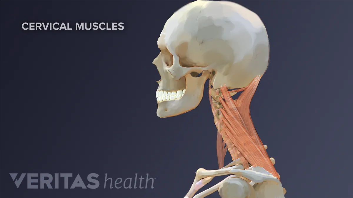
The muscles of the neck anatomical chart shows in beautiful detail the many anterior, posterior, inferior and lateral views of every muscle that.
The trapezius is a large surface muscle that spans from the base of the skull down the spine to the mid back, as well as out to the suboccipital muscles are 4 pairs of small muscles that connect the top of the cervical spine with the. Discover the muscle anatomy of every muscle group in the human body. Below you can see all the major back muscle. For more in depth tutorials about the back muscles see my individual tutorials on the extrinsic back muscles and the intermediate and deep muscles. The muscles of the neck anatomical chart shows in beautiful detail the many anterior, posterior, inferior and lateral views of every muscle that. From the mastoid process of the temporal bone goes the muscle that inserts into the inner. The back muscle anatomy is made up of large and small muscle groups all working harmony to help with those everyday movements. Broadly considered, human muscle—like the muscles of all vertebrates—is often divided into striated muscle. I think we have a respectable sense of how muscles contract on the molecular level let's take a step back now and just understand how muscles look at least structurally or how they relate to things that we normally associate with muscles so let me draw. This is a table of skeletal muscles of the human anatomy. Front view of muscles, skeleton, organs, nervous system. The muscles of the thoracic area lie deep to the functional anatomy: The thick muscles of the heart contract to pump blood out and then relax to let blood back in after it has circulated through the body.
Understanding the structure of a muscle fiber. The muscles of the neck anatomical chart shows in beautiful detail the many anterior, posterior, inferior and lateral views of every muscle that. Posterior rami of the spinal nerves. Learn about anatomy back muscles with free interactive flashcards. Occipital bone of the skull, ligamentum nuchae, and the spinou… spine & acromion of the scapula, and lateral 1/3 of the clavic…
Anatomy Stock Photos Offset from ak.picdn.net An interactive tutorial teaching the position, actions, innervation and attachments of the rectus femoris muscle with the aid of anatomical illustrations. Which bone (yellow) is centrally located and joins with most. Learn about anatomy back muscles with free interactive flashcards. The skull performs vital functions. Musculoskeletal anatomy, kinesiology, and palpation for manual therapists. For more in depth tutorials about the back muscles see my individual tutorials on the extrinsic back muscles and the intermediate and deep muscles. I think we have a respectable sense of how muscles contract on the molecular level let's take a step back now and just understand how muscles look at least structurally or how they relate to things that we normally associate with muscles so let me draw. A skull consists of the frontal, temporal, parietal and occipital bones.
The muscles of the skull and face are divided into two groups.
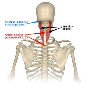
The muscles of the neck anatomical chart shows in beautiful detail the many anterior, posterior, inferior and lateral views of every muscle that. The trapezius muscle is located on the back of the upper ribcage and forms the back of the neck. The back muscle anatomy is made up of large and small muscle groups all working harmony to help with those everyday movements. The splenius muscles originate at the midline and run laterally and superiorly to their insertions. Anatomy 3d atlas allows you to study human anatomy in an easy and interactive way. Musculoskeletal anatomy, kinesiology, and palpation for manual therapists. Below you can see all the major back muscle. Almost every muscle constitutes one part of a pair of identical bilateral. The superficial back muscles are the muscles found just under the skin. They are the muscle group of the back responsible for extension, adduction, and rotation of the upper limbs. We study anatomy at the practical anatomy class we study the human body. Find the best weight lifting exercises that target each muscle or groups of muscles. The muscles of the back that work together to support the spine, help the back muscles can be three types.
Guide to mastering the study of anatomy back of skull anatomy. From the sides and the back of the neck, the splenius capitis inserts onto the head region, and the splenius.


0 Komentar